Schizophrenia Neurology: How Does It Affect the Brain?
Beyond a simple chemical imbalance: what genes, synapses, dopamine, glutamate, inhibitory circuits, and brain networks reveal about schizophrenia—and what those findings mean for care.
In this article
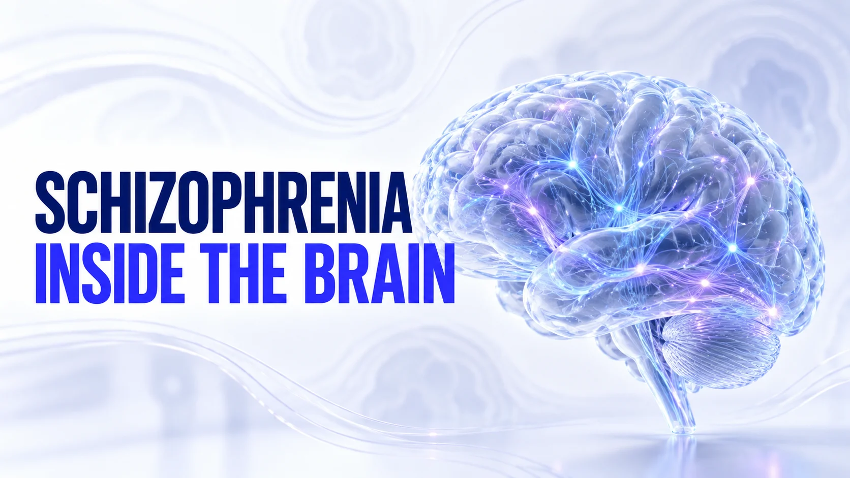
Schizophrenia changes how a person experiences and interprets the world, but it does not have one known cause or a single damaged brain region. Research is building a more detailed picture: differences in brain development and synaptic signaling may affect how networks coordinate perception, memory, motivation, and thought. Understanding that biology can make the illness less mysterious without reducing a person to a scan or a diagnosis.
Start with the symptoms biology needs to explain
Schizophrenia involves more than hallucinations. Psychotic symptoms can include hallucinations, delusions, and disorganized thinking. Negative symptoms involve reductions in motivation, expression, or engagement. Cognitive symptoms can affect attention, working memory, and decision-making. These dimensions help explain why symptom relief and day-to-day recovery are related but not identical goals.1
It is not ‘split personality,’ and reduced expression does not mean someone has no feelings. A useful biological explanation must account for this range of experiences, rather than treating every difficulty as another form of psychosis.1
A developmental vulnerability—not one schizophrenia gene
A major genetic study published in 2022 identified associations at 287 genomic locations. The implicated biology was concentrated in neurons, including excitatory and inhibitory cells, and processes involved in synaptic organization and transmission. This points toward how brain cells develop and communicate—not a single faulty chemical or gene.2
Most common risk variants have small individual effects. Their combined influence can alter vulnerability, but a risk association does not tell us that every carrier will develop schizophrenia. Nor does identifying a genetic association automatically reveal exactly how that variant changes a person's brain.2
The developmental model helps frame a key question: how might an early vulnerability become clinically apparent later? Schizophrenia often emerges in adolescence or early adulthood, although changes in thinking or functioning can precede a first psychotic episode. Genes interact with life circumstances; prenatal adversity, stressful environments, and other exposures may contribute to risk rather than determining an inevitable outcome.1
Synapses and pruning: where development meets immune biology
Synapses are the contact points through which neurons communicate. During development, connections are formed, strengthened, and selectively removed. Researchers are investigating whether altered regulation of these processes helps explain some vulnerability to schizophrenia.23
An influential study connected variation in complement component 4, particularly predicted C4A expression, with schizophrenia risk. Complement proteins belong to the immune system, but they also have functions in the nervous system. The study combined human genetic findings with experiments showing that C4 contributes to developmental synapse elimination in mice.3
This supports a possible link between immune-related biology and excessive or mistimed pruning. It does not establish that everyone with schizophrenia has the same pruning problem, that the illness is universally autoimmune, or that suppressing the immune system is a treatment. The proposed mechanism is more specific than ‘inflammation damages the brain.’3
Living-brain imaging offers another piece of evidence. A small PET study found lower binding to SV2A, a protein used as a marker of synaptic terminals, in frontal and anterior cingulate regions in schizophrenia. There were 18 participants in each group, so the finding is informative rather than a diagnostic yardstick.4
SV2A imaging does not count every synapse directly or prove that pruning caused the difference. It does, however, connect the synapse hypothesis with a measurable signal in living people, rather than relying only on genetics or tissue studied after death.4
Dopamine: a strong finding, but not ‘too much everywhere’
Dopamine is important in learning, motivation, and the significance assigned to events. One influential prospective PET study found greater dopamine synthesis capacity in the striatum among high-risk participants who later developed psychosis. The subgroup that transitioned was small, but the result showed that the difference could precede an established psychotic illness.5
The anatomical detail matters: evidence about striatal dopamine is not evidence of a uniform dopamine excess across the brain. Synthesis capacity is also not the same measurement as receptor number or dopamine concentration at every moment.5
One explanatory model is that dysregulated dopamine signaling helps ordinary events acquire unusual importance, making unexpected interpretations feel compelling. That is an interpretation of how biology might connect with experience, not a complete explanation of every delusion, hallucination, or cognitive symptom. The imaging finding alone does not prove that sequence.5
Glutamate and NMDA receptors: why excitation is not a simple volume knob
Glutamate supports excitatory communication, and NMDA receptors help regulate learning and plasticity. Interest in NMDA dysfunction partly comes from experiments in which ketamine, an NMDA receptor antagonist, produced transient perceptual, thought, and cognitive changes in healthy volunteers.6
That makes NMDA-related signaling a useful experimental route into symptoms that dopamine alone does not explain. It does not mean schizophrenia is equivalent to ketamine exposure, or that a drug challenge establishes the cause of a lifelong condition.6
A leading circuit model asks what happens if excitatory input to inhibitory neurons is weakened: less recruitment of inhibition could allow other neurons to become less precisely regulated. In awake rats, NMDA receptor blockade reduced activity in putative inhibitory interneurons while increasing activity in many pyramidal neurons. Thus, reduced function at one receptor can increase activity elsewhere. Translating this experimental result into an explanation of human schizophrenia remains a hypothesis—not a claim that all glutamate is high or low in every person.13
GABA interneurons: the timing system between signals
Inhibition does more than reduce activity; it helps organize when groups of neurons fire. Research on prefrontal GABA neurons, including cells that express parvalbumin, is relevant because cognitive tasks depend on coordinated activity rather than the sheer amount of signaling.7
Postmortem research found reduced expression of GAD67, an enzyme involved in making GABA, within a subset of these neurons. That is evidence of altered cellular signaling machinery, not proof that all inhibitory neurons are missing or that a person's brain simply needs a GABA supplement.7
The proposed consequence is less precise coordination between excitation and inhibition. Researchers study how this could contribute to difficulty maintaining information and organizing thought. Tissue findings cannot establish every step of that pathway or tell us which change came first.7
Brain networks: altered coordination, not just weaker connections
Resting-state functional MRI measures relationships between fluctuations in brain signals. In schizophrenia, some connections appear stronger and others weaker, so ‘less connectivity’ is an incomplete description. A thalamic connectivity study found altered patterns involving cortical systems and also examined similarities and differences with bipolar disorder.8
The thalamus participates in loops linking sensory processing with higher-order cortical activity. A prominent pattern is relatively reduced coupling with prefrontal systems alongside increased coupling with sensory-motor areas. This suggests altered coordination across systems, rather than one isolated malfunctioning region.8
Functional connectivity is a statistical relationship between signals, not a direct photograph of an axon or proof of which region caused a symptom. Network models are valuable because perception, attention, memory, and thought require regions to work together—but a network image cannot read someone's thoughts or confirm a diagnosis.8
What structural brain scans show—and what they do not
An ENIGMA analysis combining data from 4,474 people with schizophrenia and 5,098 comparison participants found widespread average differences in cortical thickness and surface area. Frontal and temporal regions were prominent in the thickness findings.9
Those are group averages with substantial overlap, not a required appearance of an individual brain. MRI thickness also does not directly measure neuron loss. Development, illness-related processes, treatment exposure, and other influences complicate interpretation; a cross-sectional comparison cannot separate their contributions cleanly.9
The practical distinction is important: researchers can detect statistical patterns across thousands of scans without having a scan that identifies schizophrenia in one person. Imaging may still be appropriate to investigate another suspected cause of symptoms; it is not a substitute for a clinical assessment.912
How these findings might fit together
A working synthesis is that developmental vulnerability affects synapses and inhibitory-excitatory coordination; altered network activity may then interact with dopamine systems and the interpretation of experience. This joins several lines of research into a plausible model—not a proven sequence that applies to everyone.23578
Different people may reach similar symptoms through partly different pathways. That helps explain why a discovery can be biologically important without immediately becoming a universal diagnostic test or a treatment for every aspect of schizophrenia. These models need to explain differences between people as well as what they share.
Diagnosis still requires the whole clinical picture
Assessment considers symptoms over time, functioning, medications and substances, physical health, and other possible explanations. Mood disorders, substance-related states, and medical causes of psychosis need consideration. Blood tests or scans can answer particular clinical questions, but there is no single routine biomarker that confirms schizophrenia.12
Research findings should guide better questions, not encourage self-diagnosis from a genetic score, a scan description, or an unusual experience. A timely assessment is useful precisely because several treatable problems can resemble one another.12
What brain biology means for treatment today
Many established antipsychotics act through dopamine D2-related mechanisms, but that is no longer the only approved pharmacological approach. In 2024, the FDA approved xanomeline–trospium (Cobenfy) for schizophrenia in adults, introducing an approach centered on cholinergic rather than direct dopamine-receptor targeting.11
Xanomeline acts at muscarinic acetylcholine receptors. Trospium helps reduce peripheral effects of muscarinic stimulation. This expands treatment biology, but it does not prove that a single acetylcholine deficit causes schizophrenia or remove the need to weigh side effects and suitability.11
Treatment choices require shared decisions and monitoring, including metabolic, movement-related, and other medication-specific risks. Clozapine is an important option when adequate trials of other antipsychotics have not helped sufficiently. Medicines should not be stopped or changed without prescriber guidance.12
Care also needs to address practical life goals. In the randomized RAISE early-treatment study, coordinated specialty care improved quality of life, symptoms, and involvement in work or school compared with usual community care for first-episode psychosis. The approach combined medication management, individual support, family education, and supported employment or education.10
That is a useful corrective to biological fatalism: finding brain differences does not mean a person's future is fixed. Effective support can change outcomes even while the precise mechanisms of the illness remain under investigation.10
The takeaway: a more detailed biology, not a smaller view of the person
The strongest picture is not one chemical imbalance, one gene, or one broken region. It is a developing account of synapses, signaling, and coordinated networks—with important findings and unresolved questions. Understanding that complexity can support more thoughtful care without turning research averages into predictions about an individual's life.
By Birk Gundersen RN, BSN.
Words worth knowing
Cognitive behavioral therapy (CBT)
A structured therapy that helps people notice patterns between thoughts, feelings, and actions, then practice more helpful responses.
References
- National Institute of Mental Health. Schizophrenia: symptom domains, risk factors, and care.
- Trubetskoy V, et al. Mapping genomic loci implicates genes and synaptic biology in schizophrenia. Nature. 2022;604:502–508.
- Sekar A, et al. Schizophrenia risk from complex variation of complement component 4. Nature. 2016;530:177–183.
- Onwordi EC, et al. Synaptic density marker SV2A is reduced in schizophrenia patients and unaffected by antipsychotics in rats. Nature Communications. 2020;11:246.
- Howes OD, et al. Dopamine synthesis capacity before onset of psychosis: a prospective [18F]-DOPA PET imaging study. American Journal of Psychiatry. 2011;168:1311–1317.
- Krystal JH, et al. Subanesthetic effects of the noncompetitive NMDA antagonist, ketamine, in humans. Archives of General Psychiatry. 1994;51:199–214.
- Hashimoto T, et al. Gene expression deficits in a subclass of GABA neurons in the prefrontal cortex of subjects with schizophrenia. Journal of Neuroscience. 2003;23:6315–6326.
- Anticevic A, et al. Mediodorsal and visual thalamic connectivity differ in schizophrenia and bipolar disorder with and without psychosis history. Schizophrenia Bulletin. 2014;40:1227–1243.
- van Erp TGM, et al. Cortical brain abnormalities in 4,474 individuals with schizophrenia and 5,098 controls via the ENIGMA consortium. Biological Psychiatry. 2018;84:644–654.
- Kane JM, et al. Comprehensive versus usual community care for first-episode psychosis: 2-year outcomes from the NIMH RAISE Early Treatment Program. American Journal of Psychiatry. 2016;173:362–372.
- FDA. FDA Approves Drug with New Mechanism of Action for Treatment of Schizophrenia. September 26, 2024.
- NICE. Psychosis and schizophrenia in adults: prevention and management (CG178). Assessment, shared decisions, monitoring, and treatment resistance.
- Homayoun H, Moghaddam B. NMDA receptor hypofunction produces opposite effects on prefrontal cortex interneurons and pyramidal neurons. Journal of Neuroscience. 2007;27:11496–11500. Animal electrophysiology study.